Correct Answer: A. Fibrous dysplasia
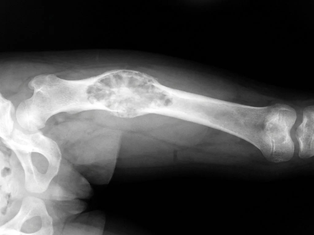
Fibrous dysplasia is a benign fibro-osseous lesion characterized by replacement of normal bone with fibrous tissue and abnormal bone formation, presenting classically with the triad of monostotic or polyostotic bone lesions, café-au-lait pigmentation, and endocrine abnormalities (McCune-Albright syndrome when polyostotic). The X-ray typically shows a "ground-glass" or "shepherd's crook" deformity in the proximal femur—a pathognomonic finding. The combination of lower limb deformity (shepherd's crook appearance from varus deformity) with hyperpigmented skin lesions (café-au-lait spots) is virtually diagnostic. Fibrous dysplasia arises from activating mutations in the GNAS gene leading to abnormal osteoblast differentiation. In Indian pediatric orthopedic practice, this is a common presentation in children aged 5–15 years. The lesion is radiolucent with ill-defined margins and may show cortical thinning. Management is conservative unless deformity is progressive; surgical correction (valgus osteotomy) is considered for severe shepherd's crook deformity to prevent pathological fractures and functional impairment.
Why the other options are wrong
B. Paget's disease — Paget's disease presents with lytic and sclerotic phases on X-ray (not ground-glass appearance) and occurs in elderly patients (>40 years), not children. It is rare in India and does not present with café-au-lait spots. The deformity is typically anterior bowing of the tibia (saber shin), not shepherd's crook. Serum alkaline phosphatase is markedly elevated, and there is no skin pigmentation association. C. Osteogenesis imperfecta — Osteogenesis imperfecta is a collagen synthesis disorder presenting with multiple fractures, blue sclerae, and hearing loss—not café-au-lait spots. X-rays show osteopenia with thin cortices and wormian bones, not ground-glass lesions. The deformity results from repeated fractures, not primary bone dysplasia. Hyperpigmentation is not a feature; this is a connective tissue disorder, not a fibro-osseous lesion. D. Non-ossifying fibroma — Non-ossifying fibroma is a small, asymptomatic, self-limiting lesion in the metaphysis of long bones (especially distal femur and tibia) that typically regresses spontaneously by age 20. It does not cause significant deformity or systemic manifestations. Crucially, it is not associated with café-au-lait spots or endocrine abnormalities. X-ray shows a well-defined radiolucent lesion with sclerotic borders, not ground-glass appearance.
High-Yield Facts
- Fibrous dysplasia triad: polyostotic lesions + café-au-lait spots + endocrine dysfunction (McCune-Albright syndrome)
- Shepherd's crook deformity: varus deformity of proximal femur pathognomonic for fibrous dysplasia on X-ray
- Ground-glass appearance: radiolucent lesion with ill-defined margins on X-ray, caused by abnormal fibrous tissue and woven bone
- GNAS mutation: activating somatic mutation in osteoblasts leads to abnormal bone formation and pigmentation
- Age of presentation: typically 5–15 years in children; monostotic form more common than polyostotic
- Management: conservative observation; valgus osteotomy for progressive deformity to prevent pathological fractures
Mnemonics
FD-CAB Fibrous dysplasia → Café-au-lait spots, Abnormal bone (ground-glass), Bone deformity (shepherd's crook). Use this to recall the classic triad when you see a child with lower limb deformity and skin pigmentation. McCune-Albright = Polyostotic FD Multiple bones + Café-au-lait + Andocrine (precocious puberty, hyperthyroidism). Remember: monostotic FD has no skin lesions; polyostotic FD = McCune-Albright syndrome. Use when differentiating extent of disease.
NBE Trap
NBE pairs café-au-lait spots with neurofibromatosis (NF-1) to lure students into missing the fibrous dysplasia + shepherd's crook combination. While both have pigmentation, only fibrous dysplasia causes characteristic bone deformity with ground-glass X-ray appearance in children.
Clinical Pearl
In Indian pediatric orthopedic clinics, fibrous dysplasia is often diagnosed late because the shepherd's crook deformity is mistaken for rickets or other metabolic bone disease. The key discriminator is the ground-glass X-ray appearance + café-au-lait spots—rickets shows metaphyseal widening and osteopenia, not ground-glass lesions. Early recognition prevents pathological fractures and guides surgical planning.
_Reference: Robbins & Cotran Pathologic Basis of Disease, Ch. 26 (Bones); Bailey & Love's Short Practice of Surgery, Ch. 39 (Orthopedics)_