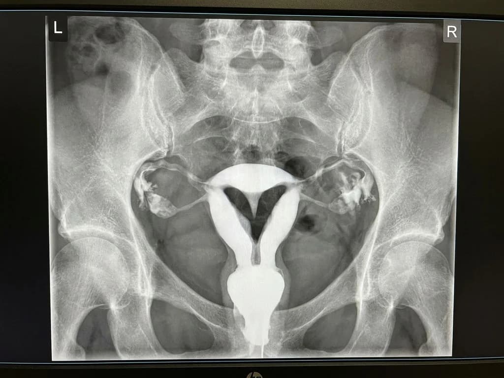
Correct Answer: B. Septate uterus
A septate uterus is the most common congenital uterine anomaly (35% of all Müllerian anomalies) and is characterized by a normal external uterine contour with an internal fibrous or muscular septum dividing the uterine cavity. On hysterosalpingography (HSG), the key discriminating feature is a normal external fundal contour (no indentation) combined with an internal septum that partially or completely divides the endometrial cavity. The septum is typically avascular and composed of fibrous tissue with poor blood supply, making it the most common anomaly associated with reproductive failure—recurrent miscarriage (40–60%), preterm labor, and intrauterine growth restriction. Unlike other Müllerian anomalies, septate uterus is the only one where hysteroscopic septum resection (metroplasty) significantly improves fertility outcomes, making accurate diagnosis critical. The external contour remains normal because both Müllerian ducts fused normally; the defect lies in incomplete resorption of the median septum during embryogenesis. HSG shows a single cervix, single external os, and a normal-appearing fundus on anteroposterior view, distinguishing it from bicornuate (which shows external indentation) and unicornuate (which shows asymmetry) variants.
Why the other options are wrong
A. Unicornuate uterus — A unicornuate uterus results from complete agenesis or severe hypoplasia of one Müllerian duct, producing an asymmetrical, banana-shaped uterus with a single fallopian tube on the developed side. HSG would show a small, eccentrically positioned uterine cavity, not the symmetric cavity with internal septum seen in septate uterus. Unicornuate uterus is associated with renal agenesis on the ipsilateral side (30–40% of cases). The external contour is distinctly asymmetrical, unlike the normal external fundal contour in septate uterus. C. Bicornuate uterus — Bicornuate uterus results from incomplete fusion of the Müllerian ducts at the fundus, creating two separate uterine horns with a normal external indentation (heart-shaped or bicornuate appearance on anteroposterior HSG view). The key difference from septate uterus is the external fundal indentation (>1 cm), which is absent in septate uterus. Both have two separate cavities, but bicornuate shows external deformity while septate shows normal external contour. Bicornuate uterus has better fertility outcomes than septate uterus and does not benefit from surgical intervention. D. Uterine didelphys — Uterine didelphys (complete duplication) results from complete failure of Müllerian duct fusion, producing two completely separate uteri, two cervices, and often a longitudinal vaginal septum. HSG would show two completely separate uterine cavities with two distinct cervical canals, not a single cavity divided by a septum. This is the rarest Müllerian anomaly (0.1–0.3% of women) and is associated with renal and skeletal anomalies. The presence of two distinct uteri on imaging immediately excludes septate uterus.
High-Yield Facts
- Septate uterus is the most common congenital uterine anomaly (35% of Müllerian anomalies) and the only one where hysteroscopic metroplasty improves fertility.
- Normal external fundal contour on HSG or 3D ultrasound is the discriminating feature that distinguishes septate from bicornuate uterus.
- Recurrent miscarriage (40–60%) and preterm labor are the hallmark reproductive complications of septate uterus due to poor blood supply to the avascular septum.
- Hysteroscopic septum resection is the definitive treatment for symptomatic septate uterus; metroplasty is NOT indicated for bicornuate or unicornuate variants.
- 3D ultrasound or MRI is superior to HSG alone for diagnosing septate uterus; HSG may underestimate the extent of the septum.
Mnemonics
SEPTATE vs BICORNUATE (External Contour Rule) Septate = Smooth external fundus (normal contour). Bicornuate = Bumpy external fundus (indentation >1 cm). Use this when HSG shows internal division: if external fundus is smooth → septate; if external fundus is indented → bicornuate. Müllerian Anomaly Fertility Outcomes (BEST to WORST) BAUD: Bicornuate (best fertility, no surgery), Arcuate (excellent), Unicornuate (poor), Didelphys (worst). Septate is NOT in this list because it's the ONLY anomaly where surgery (hysteroscopic metroplasty) dramatically improves outcomes.
NBE Trap
NBE often pairs septate uterus with "normal external contour" to lure students into choosing bicornuate (which also has two cavities but with external indentation). The trap is confusing the internal anatomy (both have septation) with the external morphology (only septate has normal fundus).
Clinical Pearl
In Indian clinical practice, septate uterus is the most common reason for recurrent first-trimester miscarriage in women with structurally normal karyotypes. A single HSG finding of internal septation with normal external fundus should prompt referral for hysteroscopic metroplasty before attempting further conception, as this single intervention can reduce miscarriage rates from 60% to <20% in subsequent pregnancies.
_Reference: DC Dutta's Textbook of Obstetrics (Congenital Anomalies of Uterus, Ch. 3); Harrison Ch. 246 (Reproductive Endocrinology)_