Correct Answer: A. Vesicoureteric reflux
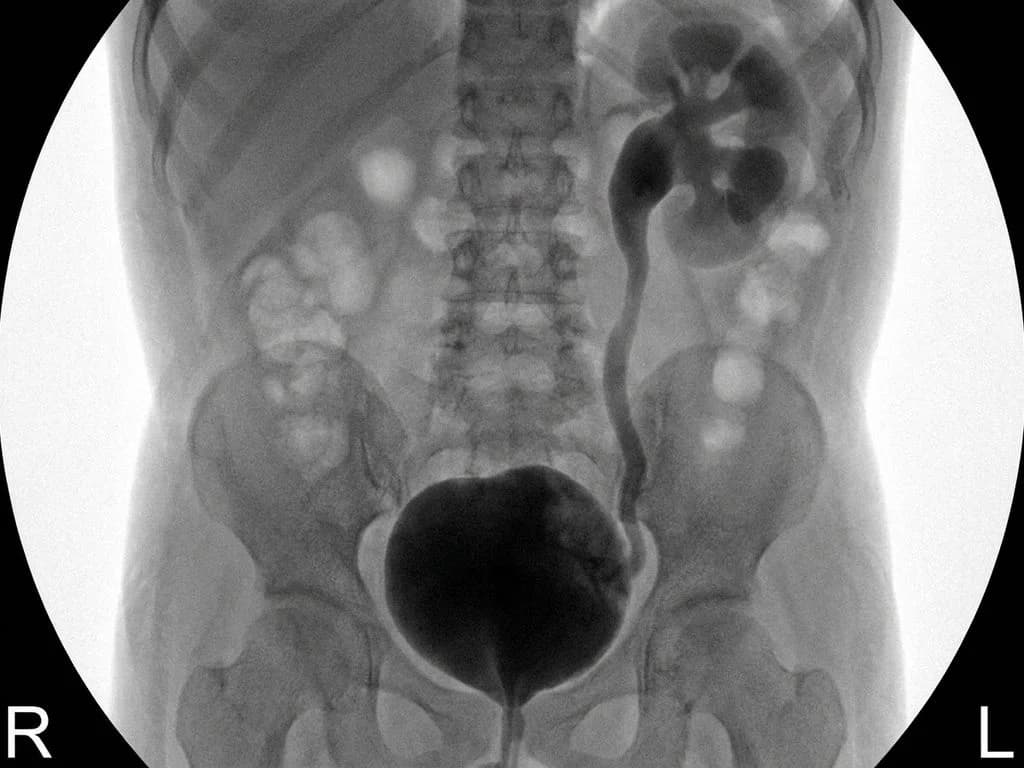
Vesicoureteric reflux (VUR) is the retrograde flow of urine from the bladder into the ureters and upper urinary tract, classically presenting with recurrent UTIs in children. The imaging (likely a voiding cystourethrogram or VCUG) would show contrast refluxing up the ureter(s) during micturition—the pathognomonic finding. In a 6-year-old boy with recurrent UTIs, VUR is the most common anatomical abnormality of the urinary tract, affecting 1–2% of children in India. The underlying defect is incompetence of the ureterovesical junction (UVJ), either due to congenital shortness of the intramural ureter or primary weakness of the ureteral orifice. Recurrent UTIs occur because infected urine refluxes into the upper tract, seeding the kidneys and causing pyelonephritis. VUR is graded I–V by the International Reflux Study; higher grades carry risk of renal scarring and chronic kidney disease. Management depends on grade: low-grade VUR (I–II) often resolves spontaneously with prophylactic antibiotics and regular imaging; high-grade VUR (IV–V) typically requires surgical reimplantation of the ureter (ureteric reimplantation or Cohen's procedure). The imaging finding of contrast in the ureter during voiding is diagnostic.
Why the other options are wrong
B. Vesicocolic fistula — A vesicocolic fistula is an abnormal communication between the bladder and colon, typically seen in Crohn's disease, diverticulitis, or malignancy—not in a 6-year-old with recurrent UTIs. Imaging would show contrast entering the colon during cystography, and the clinical presentation would include pneumaturia and fecaluria, not isolated UTIs. This is a rare condition in pediatric practice and unrelated to the UVJ incompetence causing reflux. C. Urinary bladder diverticulum — A bladder diverticulum is an outpouching of the bladder wall, which may be congenital (often at the urachal remnant or lateral wall) or acquired. While diverticula can predispose to UTIs due to urine stasis, imaging would show a discrete pouch arising from the bladder wall, not contrast refluxing into the ureters. Diverticula do not cause the characteristic retrograde ureteral filling seen on VCUG in VUR. D. Urinary bladder hernia — Bladder hernia is extremely rare in children and typically occurs through a defect in the bladder wall (e.g., after trauma or surgery), with herniation of bladder mucosa into adjacent structures. It would not present with recurrent UTIs as the primary complaint and would not show ureteral reflux on imaging. This is not a recognized cause of pediatric recurrent UTIs in Indian clinical practice.
High-Yield Facts
- VUR prevalence in children with UTI: 30–50% of children presenting with febrile UTI have VUR; it is the most common urological abnormality in pediatric practice.
- VUR grading (I–V): Grade I–II (low) often resolves spontaneously; Grade IV–V (high) requires surgical reimplantation to prevent renal scarring.
- Diagnostic imaging: VCUG (voiding cystourethrogram) is the gold standard; contrast reflux into the ureter during micturition is pathognomonic.
- Mechanism: Incompetence of the ureterovesical junction (UVJ) due to congenital shortness of the intramural ureter or primary ureteral orifice weakness.
- Renal scarring risk: Reflux of infected urine causes pyelonephritis; repeated episodes lead to renal scarring, hypertension, and CKD in 10–15% of untreated cases.
- Management: Low-grade VUR: prophylactic antibiotics (nitrofurantoin or trimethoprim-sulfamethoxazole) + regular imaging; High-grade VUR: ureteric reimplantation (Cohen's procedure or extravesical approach).
Mnemonics
VUR Presentation in Kids (REFLECT) Recurrent UTI, Elevated creatinine (if scarred), Febrile episodes, Low-grade reflux (often asymptomatic), Excretion imaging shows reflux, Chronic kidney disease (late), Treatment depends on grade. Use this to remember that VUR is the reflex diagnosis in any child with recurrent UTI. VUR Grading Memory Hook I–II = Ureter only (low-grade, often resolves), III = Ureter + renal pelvis (moderate), IV–V = Dilated system (high-grade, needs surgery). Think: higher number = higher dilation = higher surgery risk.
NBE Trap
NBE may present a case of bladder diverticulum or fistula with UTI history to trap students who confuse any bladder pathology causing UTI with VUR. The key discriminator is the imaging finding of contrast refluxing into the ureter during micturition—this is pathognomonic for VUR and not seen in diverticula or fistulae.
Clinical Pearl
In Indian pediatric practice, VUR is the leading cause of recurrent UTI in children under 10 years. Early diagnosis via VCUG and appropriate grading prevents renal scarring—a major cause of hypertension and CKD in Indian children. Prophylactic antibiotics and regular follow-up imaging are cost-effective alternatives to early surgery in low-grade VUR.
_Reference: Bailey & Love Ch. 76 (Urology); OP Ghai Ch. 18 (Pediatric Surgery); Harrison Ch. 279 (Urinary Tract Infections)_