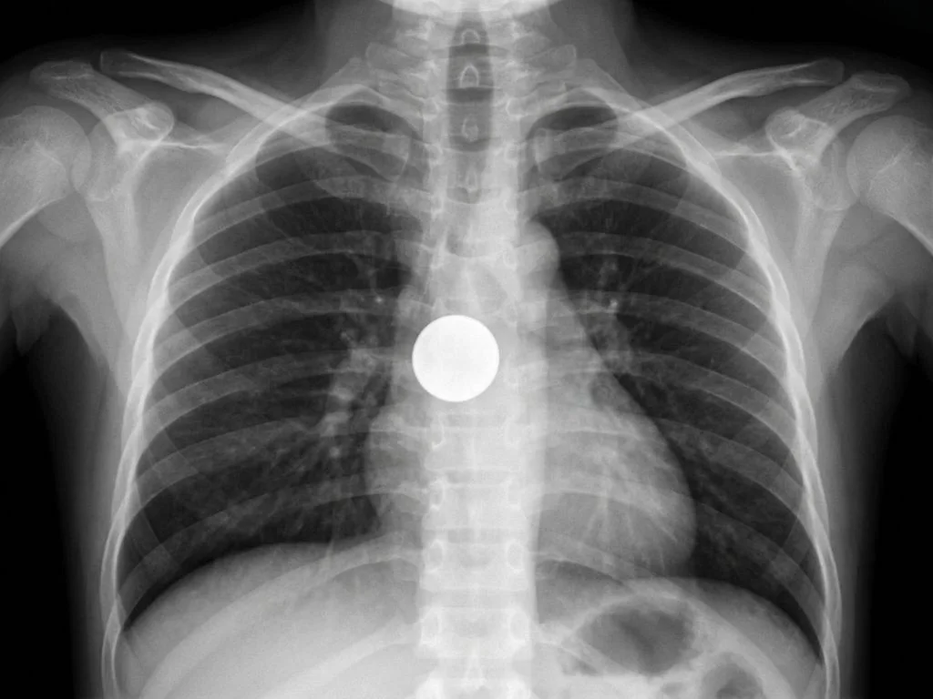
Correct Answer: B. Foreign body in the esophagus
The clinical presentation of acute dysphagia in an unsupervised child with a radiopaque object on X-ray is pathognomonic for esophageal foreign body. The key discriminating feature is the location and orientation of the radiopaque object on the anteroposterior (AP) chest X-ray. In esophageal foreign bodies, the object typically appears in the midline or slightly off-midline on AP view, and on lateral view it projects within the esophageal lumen (which lies posterior to the trachea). The esophagus is a muscular tube that runs vertically from the pharynx (C6 level) to the gastroesophageal junction (T11 level), and foreign bodies lodge most commonly at the cricopharyngeus muscle (upper esophageal sphincter), the aortic arch level (mid-esophagus), or the lower esophageal sphincter. In Indian pediatric practice, common esophageal foreign bodies include button batteries, coins, and food boluses. The acute onset of dysphagia without respiratory distress is the clinical clue that distinguishes esophageal from tracheal lodgment. Management involves endoscopic removal under general anesthesia, with NPO status and IV fluids initiated immediately to prevent aspiration and esophageal perforation.
Why the other options are wrong
A. Artifact — This is wrong because the radiopaque object on X-ray shows consistent density and defined borders, which are features of a true foreign body, not an artifact. Artifacts typically appear as linear streaks, beam hardening, or ill-defined shadows. The clinical correlation of acute dysphagia in an unsupervised child makes artifact diagnosis untenable—NBE expects recognition that clinical history + imaging findings together confirm pathology. C. Foreign body in the trachea — This is wrong because tracheal foreign bodies present with acute respiratory distress, stridor, or cyanosis—not isolated dysphagia. On X-ray, tracheal objects appear in the midline on AP view and project anteriorly on lateral view (anterior to the esophagus). The absence of respiratory symptoms and the posterior/esophageal location of the object rule out tracheal lodgment. NBE pairs these two conditions to trap students who confuse anatomical location with clinical presentation. D. Soft tissue calcification in the neck — This is wrong because soft tissue calcifications are chronic findings (seen in tuberculosis, sarcoidosis, or dystrophic calcification) and present with no acute symptoms. The clinical presentation of acute dysphagia in an unsupervised child rules out this diagnosis. Calcifications also show a diffuse, granular pattern rather than the discrete, well-defined radiopacity of a foreign body. This is a distractor for students who see any radiopaque finding and default to calcification.
High-Yield Facts
- Cricopharyngeus muscle (C6 level) is the most common site of esophageal foreign body lodgment in children.
- Button batteries are the most dangerous esophageal foreign bodies in India—cause transmural necrosis within 2–4 hours; require emergent endoscopic removal.
- Dysphagia without respiratory distress is the clinical red flag for esophageal (not tracheal) foreign body.
- AP and lateral chest X-rays are mandatory to localize the foreign body; radiopaque objects (coins, batteries) are visible; radiolucent objects (food, plastic) require contrast studies or endoscopy.
- NPO status and IV fluids are initiated immediately; endoscopic removal under GA is the gold standard within 6–12 hours to prevent esophageal perforation and mediastinitis.
Mnemonics
**SAFE Foreign Body Removal Supervision (ensure NPO, IV access, GA readiness) | Assessment (AP + lateral X-rays, endoscopy) | Foreign body removal (endoscopic, within 6–12 hours) | Examine for perforation (post-removal imaging, observe for fever/chest pain). Dysphagia = Esophagus; Stridor = Trachea** Isolated dysphagia → esophageal FB. Stridor, cyanosis, respiratory distress → tracheal FB. This 5-second rule separates the two in exams.
NBE Trap
NBE pairs esophageal and tracheal foreign bodies to trap students who confuse anatomical location with clinical presentation. Students may select tracheal FB if they see a radiopaque object without carefully reading the dysphagia-only symptom. The absence of respiratory distress is the key discriminator.
Clinical Pearl
In Indian emergency departments, unsupervised toddlers presenting with acute dysphagia after playing should trigger immediate X-ray and NPO status. Button batteries are increasingly common in urban India—any radiopaque object in the esophagus warrants same-day endoscopic removal to prevent catastrophic mediastinitis.
_Reference: Bailey & Love Ch. 62 (Esophagus); OP Ghai Ch. 9 (Pediatric Surgery)_
