Correct Answer: B. Brodie's abscess
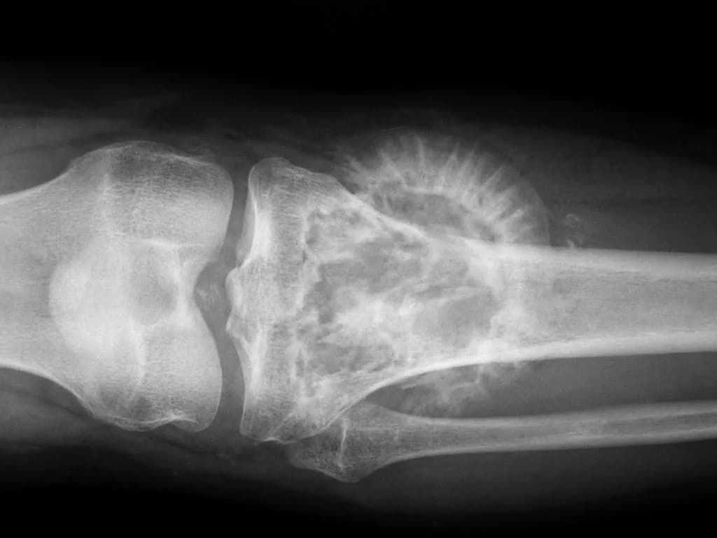
Brodie's abscess is a localized, chronic osteomyelitis that presents with a well-demarcated, sclerotic lesion typically in the metaphyseal region of long bones—classically the upper tibia in children. The 6-month gradual progression with pain and swelling, combined with the radiographic appearance of a small, round, sclerotic lesion with a surrounding zone of sclerosis (the pathognomonic "nidus" surrounded by reactive bone), is diagnostic. Unlike acute osteomyelitis, Brodie's abscess develops insidiously, often following a subclinical or inadequately treated infection. The lesion represents a localized collection of pus walled off by fibrous tissue and new bone formation. The upper tibia is the most common site (>50% of cases), followed by the distal femur. In Indian pediatric populations, this is typically seen as a sequela of hematogenous spread or direct inoculation. The sclerotic margin on radiography distinguishes it from acute osteomyelitis (which shows ill-defined margins) and from neoplastic lesions. Treatment involves surgical drainage and curettage, often combined with antibiotics, particularly if culture yields organisms like Staphylococcus aureus.
Why the other options are wrong
A. Osteoblastoma — Osteoblastoma is a benign bone tumor that presents with pain relieved by NSAIDs (unlike Brodie's abscess) and shows a larger, less sclerotic lesion without the characteristic nidus-and-sclerosis pattern. It typically occurs in the posterior elements of vertebrae or femur, not the metaphyseal tibia. The 6-month duration with progressive swelling is more consistent with infection than neoplasia. C. Ewing's sarcoma — Ewing's sarcoma is a malignant tumor presenting with rapid onset (weeks to months), systemic symptoms (fever, malaise), and an ill-defined, permeative lesion with onion-skin periosteal reaction on radiography. It typically affects the diaphysis or diaphyseal-metaphyseal region, not the metaphysis alone. The well-demarcated sclerotic lesion with nidus is incompatible with Ewing's aggressive radiographic pattern. D. Osteosarcoma — Osteosarcoma is an aggressive malignancy with rapid progression, cortical destruction, and a sunburst or Codman's triangle periosteal reaction. It occurs in adolescents around the knee (distal femur or proximal tibia) but shows a large, destructive lesion with mixed lytic-sclerotic areas, not the small, well-circumscribed nidus typical of Brodie's abscess. Systemic symptoms and rapid deterioration are expected.
High-Yield Facts
- Brodie's abscess = chronic localized osteomyelitis with pathognomonic nidus surrounded by sclerotic margin on radiography.
- Upper tibia metaphysis is the most common site (>50%), followed by distal femur; typically in children aged 5–15 years.
- 6-month insidious progression with pain and swelling (no acute systemic toxicity) is the clinical hallmark; distinguishes it from acute osteomyelitis.
- Staphylococcus aureus is the most common causative organism; culture from surgical drainage confirms diagnosis.
- Treatment = surgical drainage and curettage ± antibiotics; imaging shows complete resolution of the lesion post-operatively.
Mnemonics
Brodie's = Boring Bone Infection Brodie's = Boring (chronic, slow), Bone (osteomyelitis), Incapsulated (sclerotic wall), Nidus (central pus collection). Contrasts with acute osteomyelitis which is aggressive and ill-defined. Brodie's Site: TiFe (Tibia-Femur) Tibia (upper metaphysis, >50%) and Femur (distal, second most common). Both around the knee joint in children.
NBE Trap
NBE pairs the metaphyseal location and sclerotic appearance with benign tumors (osteoblastoma) or aggressive malignancies (Ewing's, osteosarcoma) to trap students who confuse chronic infection with neoplasia. The key discriminator is the nidus-and-sclerosis pattern plus the 6-month indolent course in a child—classic for Brodie's, not tumors.
Clinical Pearl
In Indian pediatric orthopedic practice, Brodie's abscess is often a delayed diagnosis because the insidious presentation is mistaken for a benign lesion or the child is lost to follow-up after initial antibiotic therapy for presumed acute osteomyelitis. Imaging with CT or MRI (if available) shows the central nidus and surrounding sclerosis clearly, guiding definitive surgical drainage.
_Reference: Bailey & Love Ch. 35 (Bone & Joint Infections); Robbins Ch. 26 (Bone Pathology)_