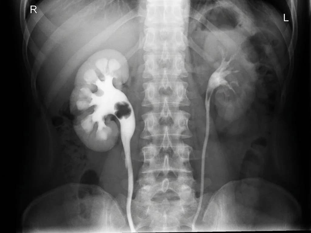
Correct Answer: D. Hydronephrosis
Hydronephrosis is the pathological dilation of the renal pelvis and calyces due to obstruction of urine flow, leading to increased intrapelvic pressure. On retrograde ureteropyelogram (RUP), the hallmark finding is progressive dilation of the collecting system — the renal pelvis appears enlarged and the calyces become blunted and dilated, with the ureter showing a transition zone where obstruction occurs. The key discriminator in RUP is that contrast fills the dilated system and outlines the site of obstruction (whether at the ureteropelvic junction, mid-ureter, or ureterovesical junction). Loin pain in hydronephrosis results from increased intrapelvic pressure and stretching of the renal capsule. The RUP image would show the characteristic "clubbed" or blunted calyces, dilated renal pelvis, and often a narrowed segment distal to the dilation — the pathognomonic appearance of obstructive uropathy. This is the most common cause of loin pain with imaging findings of collecting system dilation in Indian clinical practice, particularly from ureteric stones, UPJ obstruction, or retroperitoneal fibrosis.
Why the other options are wrong
A. Duplex kidney — Duplex kidney (complete or incomplete duplication of the collecting system) is a congenital anomaly that appears as two separate pelvicalyceal systems on RUP. However, it does NOT cause loin pain unless complicated by obstruction or reflux. The RUP would show two distinct pelvises and ureters, not the dilated collecting system with obstruction that produces acute loin pain. Duplex kidney is an anatomical variant, not an obstructive pathology. B. Renal stone — While renal stones cause loin pain and may be visible on plain radiography or CT, they are NOT the primary diagnosis here — the RUP image shows collecting system dilation (hydronephrosis), not a stone. A stone may cause hydronephrosis if it obstructs, but the radiological finding of dilated calyces and pelvis is hydronephrosis itself. The question asks for diagnosis based on the RUP appearance, not the underlying etiology. C. Renal carcinoma — Renal carcinoma typically presents with hematuria or incidental finding, not acute loin pain with hydronephrosis. On RUP, renal carcinoma would show a mass effect or distortion of the collecting system, not the smooth, progressive dilation seen in obstruction. Carcinoma does not produce the characteristic blunted calyces and dilated pelvis pattern of hydronephrosis. This is a trap for students confusing mass lesions with obstructive pathology.
High-Yield Facts
- Retrograde ureteropyelogram (RUP) shows dilated, blunted calyces and enlarged renal pelvis in hydronephrosis — the gold standard for defining the level and nature of obstruction.
- Loin pain + collecting system dilation on imaging = hydronephrosis until proven otherwise; the pain results from increased intrapelvic pressure (>20 mmHg) stretching the renal capsule.
- UPJ obstruction and ureteric stones are the two most common causes of hydronephrosis in India; RUP localizes the obstruction site precisely.
- Blunted calyces (loss of sharp angle at fornix) and dilated renal pelvis are pathognomonic RUP findings of chronic hydronephrosis.
- Duplex kidney is a congenital anomaly with two pelvices but does NOT cause loin pain or dilation unless obstructed — it is an anatomical finding, not a diagnosis of obstruction.
Mnemonics
HYD-RO (Hydronephrosis Recognition on Imaging) High intrapelvic pressure → Yield dilated calyces → Dilated pelvis → Retrograde shows obstruction → Obstruction site identified. Use when interpreting any RUP or ultrasound showing collecting system dilation. PAIN-DILATION Rule Pain (loin) + Acute presentation + Imaging shows dilation + Narrowing/obstruction = Diagnosis is hydronephrosis (not the stone or tumor causing it). Helps distinguish the radiological diagnosis from the etiology.
NBE Trap
NBE pairs "loin pain" with "stone" to lure students into choosing renal stone; however, the RUP image shows the consequence of obstruction (hydronephrosis), not the stone itself. The question tests whether students can distinguish the radiological diagnosis from the underlying etiology.
Clinical Pearl
In Indian emergency departments, a patient presenting with acute loin pain and RUP showing dilated calyces is hydronephrosis until the obstruction is relieved — whether by stone passage, JJ stent, or nephrostomy. The RUP finding of dilation is the diagnosis; the stone or stricture is the cause.
_Reference: Bailey & Love Ch. 73 (Urology — Hydronephrosis and Obstruction); Harrison Ch. 279 (Urinary Tract Obstruction)_