Correct Answer: D. Great auricular nerve
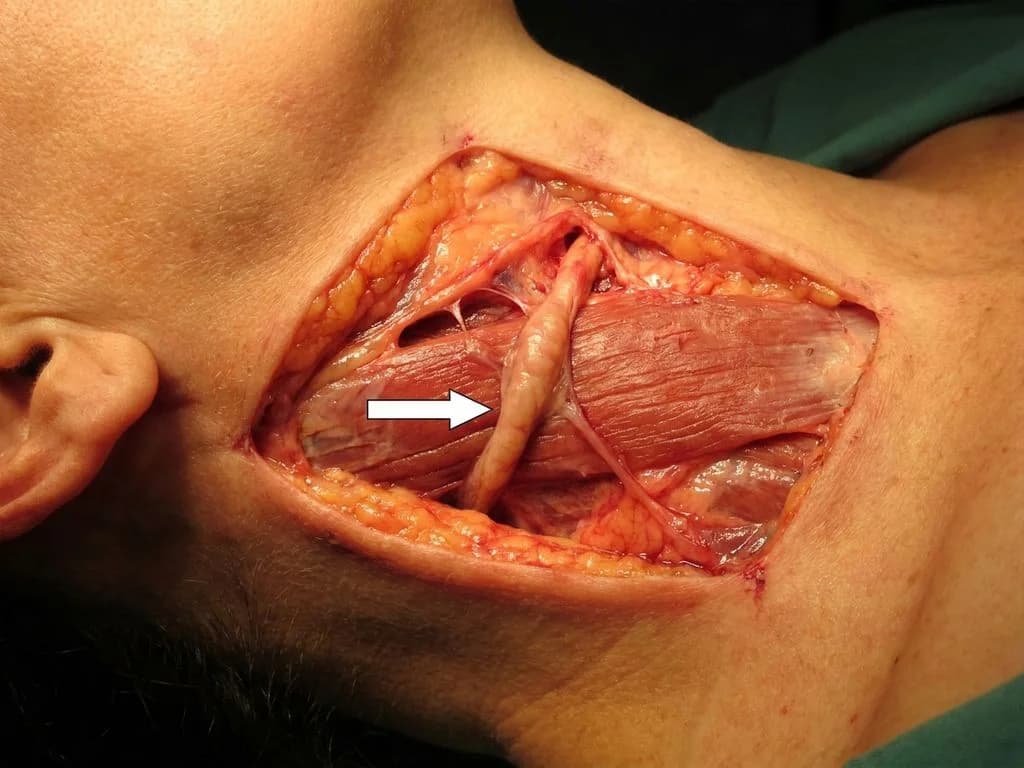
The great auricular nerve (GAN) is the largest sensory branch of the cervical plexus (C2–C3), arising from the posterior border of the sternocleidomastoid muscle. It ascends along the external jugular vein and divides into anterior and posterior branches to supply the auricle, lower part of the auricle, and skin over the angle of the mandible and parotid region. On imaging or anatomical dissection, the GAN appears as a notably thickened nerve trunk compared to other cervical sensory nerves because of its large diameter and high fiber content. This nerve is clinically significant in Indian surgical practice—particularly during parotidectomy, facelift procedures, and neck dissections—where iatrogenic injury causes numbness of the auricle and lower ear region. The GAN's prominent size and superficial course along the SCM make it easily identifiable on cross-sectional imaging and cadaveric specimens, which is why it is frequently tested in anatomy examinations. Its thickness distinguishes it from the smaller auriculotemporal (a branch of V3), lesser occipital (C2), and facial nerve proper in the neck region.
Why the other options are wrong
A. Auriculotemporal nerve — The auriculotemporal nerve is a terminal sensory branch of the mandibular division (V3) of the trigeminal nerve. It is considerably smaller in diameter than the great auricular nerve and runs within the parotid gland, not superficially along the neck. It supplies the temporal region and upper auricle, but its caliber is much thinner than the GAN. NBE may trap students who confuse auricular innervation patterns without considering nerve size and location. B. Lesser occipital nerve — The lesser occipital nerve arises from C2 (cervical plexus) and is a small sensory branch supplying the skin over the mastoid process and occipital region. It is significantly thinner than the great auricular nerve and runs more posteriorly along the posterior border of the SCM. Its small caliber and posterior course distinguish it clearly from the prominent GAN visible on imaging. C. Facial nerve — The facial nerve (CN VII) is a mixed nerve (motor + parasympathetic + sensory) that exits the stylomastoid foramen and immediately divides into its terminal branches within the parotid gland. In the neck, the facial nerve proper is not a prominent superficial structure like the GAN. The thickened nerve marked in the image is a sensory cervical plexus branch, not the facial nerve trunk.
High-Yield Facts
- Great auricular nerve (C2–C3) is the largest sensory branch of the cervical plexus and the only cervical nerve that ascends to the head.
- GAN injury during parotidectomy or facelift causes numbness of the auricle, lower ear, and angle of mandible—a common complication in Indian head and neck surgery.
- Auriculotemporal nerve (V3 branch) supplies the upper auricle and temporal region but is much smaller in diameter than GAN.
- Lesser occipital nerve (C2) is a small branch supplying mastoid and occipital skin; it runs posteriorly, not anteriorly like GAN.
- GAN anatomy: arises at posterior SCM border, ascends along external jugular vein, divides into anterior (parotid/mandibular angle) and posterior (auricle) branches.
Mnemonics
Cervical Plexus Sensory Branches (Superficial) Great auricular (C2–C3, largest, ascends to ear) | Lesser occipital (C2, posterior) | Transverse cervical (C2–C3, anterior neck) | Supraclavicular (C3–C4, shoulder). GAN is the thickest and most superior of these branches. Auricular Nerve Supply ("Two Nerves, Two Levels") Upper auricle = Auriculotemporal (V3, small, thin). Lower auricle + angle of jaw = Great auricular (C2–C3, large, thick). Remember: GAN is the big cervical nerve going up to the ear.
NBE Trap
NBE exploits confusion between the three nerves supplying the auricle (auriculotemporal, lesser occipital, and great auricular) by testing nerve size and location rather than just innervation territory. Students who only memorize "which nerve supplies which part" without visualizing anatomical thickness and course will misidentify the thickened nerve on imaging.
Clinical Pearl
In Indian head and neck surgical practice, GAN injury is the most common complication of parotidectomy and facelift procedures, causing permanent numbness of the auricle and lower ear—a complication that significantly impacts patient quality of life. Recognizing the GAN's prominent size on preoperative imaging helps surgeons plan nerve-sparing dissection.
_Reference: Clinically Oriented Anatomy by Moore & Dalley (Ch. 8: Head and Neck), Bailey & Love's Short Practice of Surgery (Ch. 40: Parotid gland and facial nerve)_