Glomerulonephritis for NEET PG — Complete Guide 2026
Master glomerulonephritis for NEET PG 2026: nephrotic vs nephritic syndromes, minimal change, FSGS, membranous, IgA, PSGN, MPGN, RPGN, lupus nephritis, Alport syndrome, and biopsy-directed management.
Version 1.0 — Published January 2026
Quick Answer
Glomerulonephritis contributes 3–4 NEET PG questions per paper. Master these 10 high-yield anchors:
- Nephrotic vs nephritic — nephrotic: proteinuria >3.5 g/24 h, hypoalbuminaemia, oedema, hyperlipidaemia, bland sediment; nephritic: haematuria with dysmorphic RBCs and RBC casts, HTN, raised creatinine
- MCD — commonest nephrotic syndrome in children (1–10 y); podocyte foot-process effacement on EM; steroid-responsive (90%)
- FSGS — focal segmental sclerosis; HIV-associated collapsing variant; APOL1 risk alleles; prolonged steroids ± CNIs/rituximab
- Membranous — anti-PLA2R (70–80% primary); spikes on silver stain; rule out malignancy, HBV, SLE; modified Ponticelli or rituximab for high-risk
- IgA nephropathy — commonest primary GN globally; synpharyngitic haematuria (within 1–3 d of URI); RAAS blockade + steroids in high-risk (KDIGO 2021)
- PSGN — 1–3 wk post-pharyngitis / 3–6 wk post-pyoderma; low C3 (recovers <8 wk); subepithelial humps on EM; supportive care
- MPGN — tram-track GBM; split into C3 glomerulopathy (alternative complement dysregulation) and immune-complex (HCV cryo, SLE)
- RPGN — crescents >50% of glomeruli; type I anti-GBM, type II immune-complex, type III pauci-immune ANCA (GPA/MPA/EGPA); pulse steroids + cyclophosphamide/rituximab ± plasmapheresis
- Lupus nephritis — ISN/RPS 2003: I minimal, II mesangial, III focal, IV diffuse, V membranous, VI advanced sclerosis; III/IV = immunosuppress
- Alport — X-linked COL4A5 (85%); GN + sensorineural deafness + lenticonus; basket-weave GBM; thin basement membrane disease is the benign familial haematuria mimic
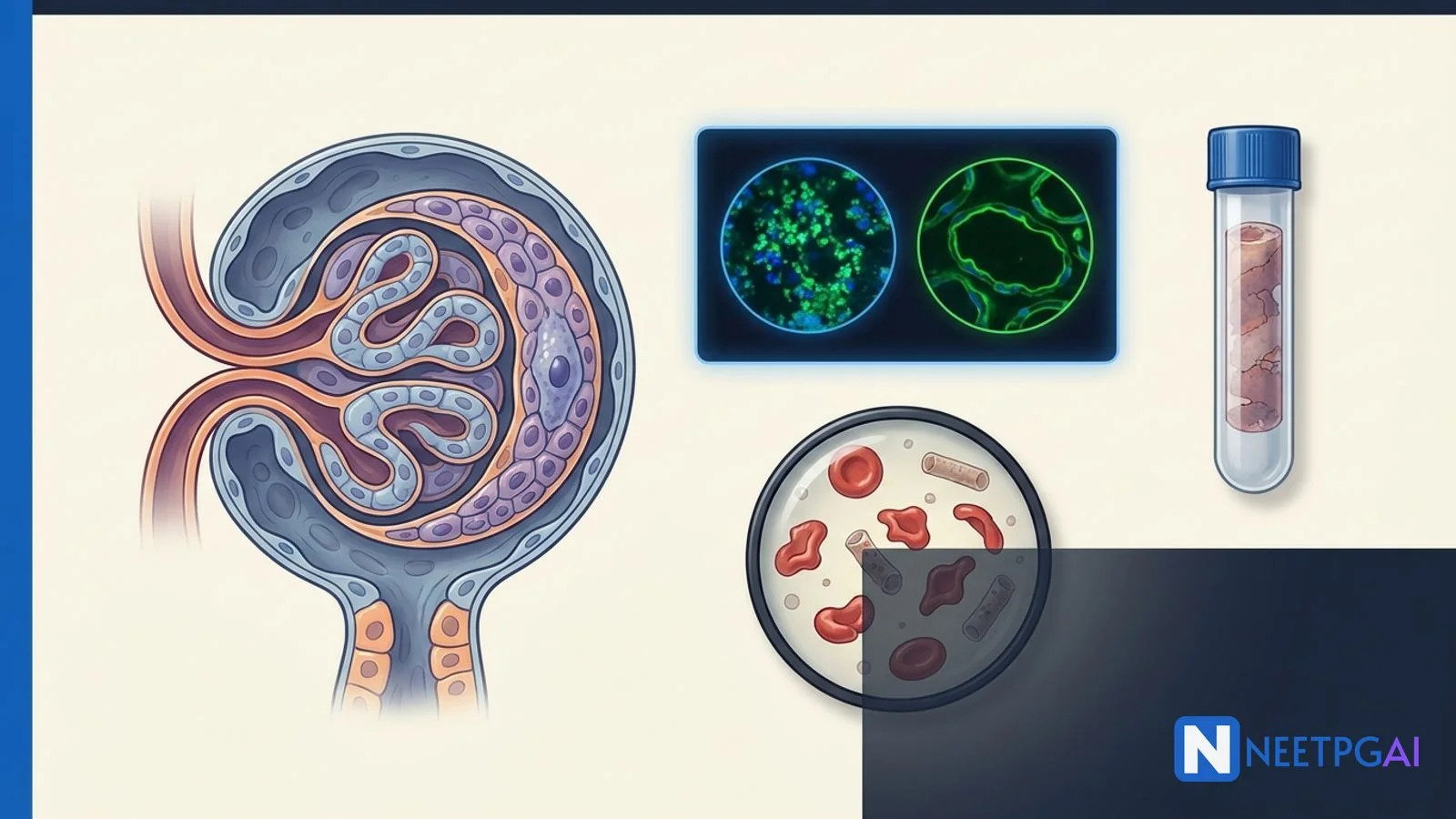
Glomerulonephritis vignettes pepper NEET PG — the child with periorbital oedema and frothy urine, the man with cola-coloured urine 2 weeks after sore throat, the young woman with malar rash and proteinuria, the smoker with haemoptysis and acute renal failure. Getting the nephrotic-vs-nephritic decision right, reading the complement pattern, and recognising the named antibodies (anti-PLA2R, ANCA, anti-GBM) separates marks. This guide covers syndromes, biopsy patterns, lupus classes, Alport, and management. Pair with the medicine subject hub, the AKI and CKD guide, and the autoimmune and connective tissue disease guide for integrated revision.
Nephrotic vs nephritic syndromes
Nephrotic and nephritic syndromes are the two clinical phenotypes of glomerular disease — defined by urinary features, mechanism of injury, and renal function — and separating them frames all further work-up.
Nephrotic syndrome (non-inflammatory podocyte / basement membrane injury):
| Feature | Value |
|---|---|
| Proteinuria | >3.5 g/24 h (adult); >40 mg/m²/h or urine protein/creatinine ratio >2 (child) |
| Hypoalbuminaemia | Serum albumin <3 g/dL |
| Oedema | Pitting, periorbital, anasarca |
| Hyperlipidaemia | Elevated LDL, TG |
| Lipiduria | Oval fat bodies, Maltese cross under polarised light |
| Sediment | Bland (few cells/casts) |
| Thrombosis risk | Renal vein thrombosis (especially membranous), DVT, PE (loss of antithrombin III) |
| Infection risk | Loss of IgG, pneumococcal spontaneous bacterial peritonitis in children |
Nephritic syndrome (inflammatory endothelial / mesangial injury):
| Feature | Value |
|---|---|
| Haematuria | Dysmorphic RBCs, RBC casts |
| Proteinuria | Variable, usually <3.5 g/24 h (overlap "nephritic-nephrotic" in severe disease) |
| Hypertension | Common |
| Oliguria and rising creatinine | Common |
| Oedema | From sodium retention |
Common causes by syndrome:
| Syndrome | Major primary causes | Major secondary causes |
|---|---|---|
| Nephrotic | MCD, FSGS, membranous nephropathy, MPGN | Diabetic nephropathy, amyloid, SLE (class V), HIV, HBV/HCV, malignancy, drugs |
| Nephritic | IgA nephropathy, PSGN, MPGN, RPGN | SLE (III/IV), vasculitis (GPA/MPA), cryoglobulinaemia, anti-GBM, endocarditis |
Complement pattern as a bedside clue:
| Low C3 / C4 | Normal complement |
|---|---|
| PSGN (low C3, normal C4) | IgA nephropathy |
| MPGN (C3 glomerulopathy: low C3; type I: low C3 + C4) | Anti-GBM |
| Lupus nephritis (low C3 and C4) | ANCA-associated (pauci-immune) |
| Cryoglobulinaemia (low C4 > C3) | Minimal change |
| Endocarditis, shunt nephritis | FSGS |
Minimal change disease and FSGS
Minimal change disease and focal segmental glomerulosclerosis sit at the primary-podocytopathy end of the nephrotic spectrum — both cause foot-process effacement, but light microscopy and prognosis differ.
Minimal change disease (MCD):
- Commonest cause of nephrotic syndrome in children (~80–90%, peak 2–6 years)
- 10–15% of adult nephrotic syndrome
- Light microscopy — normal glomeruli
- Immunofluorescence — negative
- Electron microscopy — diffuse podocyte foot-process effacement (pathognomonic)
- Associations — NSAIDs, Hodgkin lymphoma, allergy, viral
- Management (paediatric):
- Presumptive diagnosis; biopsy NOT needed in typical 1–10 y child
- Oral prednisolone 60 mg/m²/day × 4–6 weeks, then tapered (KDIGO 2021)
- Steroid-sensitive (~90%); frequent relapsers and steroid-dependent need calcineurin inhibitors, mycophenolate, cyclophosphamide, or rituximab
- Adults — confirmed by biopsy; slightly lower and slower response rate than children
Focal segmental glomerulosclerosis (FSGS):
- Focal = some glomeruli; segmental = part of glomerulus
- Commonest cause of adult nephrotic syndrome in many populations; increasing in incidence
- Light microscopy — focal segmental sclerosis with hyalinosis, foam cells; IF negative or non-specific (trapping)
- Variants (Columbia classification) — tip, perihilar, cellular, collapsing, NOS
| FSGS variant | Feature | Prognosis |
|---|---|---|
| Tip | Segmental lesion at tubular pole | Best prognosis; often steroid-responsive |
| Perihilar | Near vascular pole | Often secondary (adaptive) |
| Cellular | Endocapillary hypercellularity | Intermediate |
| Collapsing | Global collapse + visceral hyperplasia | Worst; HIV, parvovirus, IFN, bisphosphonates |
| NOS | Unclassified | Default |
- Primary FSGS — circulating permeability factor (suPAR, anti-nephrin); steroids + immunosuppression
- Secondary FSGS — adaptive hyperfiltration (obesity, reflux, reduced nephron mass), HIV, heroin, lithium, pamidronate, APOL1 risk alleles (African ancestry)
- Genetic FSGS — NPHS1 (nephrin), NPHS2 (podocin), ACTN4, TRPC6
- Management:
- Primary — prolonged steroids (4–6 months); steroid-resistant → CNI (tacrolimus, ciclosporin) or rituximab
- Secondary / genetic — RAAS blockade and treat cause; steroids NOT indicated
- HIV-associated nephropathy (HIVAN) — ART dramatically improves outcomes
Membranous nephropathy and IgA nephropathy
Membranous nephropathy (MN) and IgA nephropathy represent two classic primary glomerulonephritides — one nephrotic and subepithelial, one nephritic and mesangial.
Membranous nephropathy:
- Commonest primary cause of adult nephrotic syndrome in many series
- Primary (idiopathic) — ~75%; anti-PLA2R antibody in 70–80% (phospholipase A2 receptor on podocytes); anti-THSD7A in ~5%; anti-NELL1 etc. are newer antigens
- Secondary — malignancy (solid tumours in >60 y), HBV, HCV, SLE (class V), drugs (penicillamine, gold, NSAIDs, captopril), autoimmune, post-stem-cell transplant
- Light microscopy — diffuse thickening of GBM; spikes on silver (JMS) stain
- IF — diffuse granular IgG + C3 along GBM ("beaded")
- EM — subepithelial electron-dense deposits (Ehrenreich-Churg stages I–IV)
- Complications — renal vein thrombosis (highest risk in MN), pulmonary embolism
- Management:
- Risk stratification (KDIGO 2021): low / moderate / high / very-high risk by GFR, proteinuria, anti-PLA2R titre
- Low risk — RAAS blockade, diuretics, statin, anticoagulation if albumin <2 g/dL
- Moderate–high risk — immunosuppression: rituximab (first-line alternative) or modified Ponticelli (alternating monthly IV methylprednisolone + oral cyclophosphamide × 6 months)
- Very-high risk / rituximab-resistant — cyclophosphamide-based or calcineurin inhibitors
- Exclude and treat secondary cause first
IgA nephropathy (Berger disease):
- Commonest primary GN globally; high prevalence in East Asia
- Classic — synpharyngitic haematuria (episodic visible haematuria within 1–3 days of URI or mucosal infection) — distinguish from PSGN (10–21 day latency)
- Also — isolated microscopic haematuria, proteinuria, hypertension, nephrotic range in advanced disease
- Light microscopy — mesangial proliferation
- IF — dominant IgA deposits in the mesangium, often with C3
- EM — mesangial electron-dense deposits
- Oxford MEST-C score (KDIGO classification) — mesangial hypercellularity (M), endocapillary (E), segmental sclerosis (S), tubular atrophy (T), crescents (C)
- Management (KDIGO 2021):
- RAAS blockade (ACEi/ARB) for proteinuria >0.5 g/day, target BP <120/80
- Glucocorticoids (or enteric-coded budesonide targeting Peyer's patches) for persistently high-risk (proteinuria >1 g/day after 3–6 months of optimal supportive care)
- Cyclophosphamide + steroids for RPGN / crescentic IgA
- SGLT2 inhibitors (dapagliflozin, empagliflozin) reduce proteinuria and slow progression
- Henoch-Schönlein purpura (IgA vasculitis) is the systemic form — purpura, arthritis, abdominal pain, nephritis; same renal treatment principles
Post-streptococcal GN and MPGN
Post-streptococcal glomerulonephritis and membranoproliferative glomerulonephritis both cause nephritic syndrome with hypocomplementaemia — but temporal pattern, pathology, and prognosis separate them.
Post-streptococcal glomerulonephritis (PSGN):
- Classic paediatric nephritic syndrome, ages 3–12 years
- Trigger — group A beta-haemolytic streptococcus (certain "nephritogenic" M serotypes — 1, 4, 12, 49, 55)
- Latency:
- Pharyngitis → GN: 1–3 weeks
- Pyoderma → GN: 3–6 weeks
- Presentation — cola-coloured urine, periorbital oedema (morning), hypertension, oliguria, raised creatinine
- Investigations:
- ASO titre (post-pharyngitis, up in ~80%), anti-DNase B (post-pyoderma, more sensitive), streptozyme panel
- Low C3 (<50 mg/dL), normal C4 (alternative pathway activation)
- C3 recovers within 8 weeks — prolonged low C3 suggests MPGN / C3 glomerulopathy / lupus
- Urine — dysmorphic RBCs, RBC casts, proteinuria
- Biopsy (rarely needed) — diffuse endocapillary hypercellularity; IF granular "starry-sky" IgG + C3 along capillary loops; EM subepithelial humps
- Management — supportive:
- Salt and fluid restriction, loop diuretics, antihypertensives (CCB, frusemide)
- Antibiotics if infection still present (does not alter GN course but prevents spread)
- Most children recover fully; adults have worse prognosis (up to 50% with residual CKD)
Membranoproliferative glomerulonephritis (MPGN):
- Old classification — type I (subendothelial deposits, immune-complex), type II (dense deposit disease, intramembranous C3), type III (mixed)
- Modern classification by IF:
- Immune-complex MPGN — infections (HCV + cryoglobulinaemia, HBV, endocarditis, shunt nephritis, chronic infection), autoimmune (SLE, Sjögren), monoclonal gammopathy (MGRS)
- C3 glomerulopathy — alternative complement pathway dysregulation: C3 glomerulonephritis and dense deposit disease (DDD); autoantibodies (C3 nephritic factor, anti-Factor H) or genetic mutations (CFH, CFI)
- Light microscopy — lobular accentuation, mesangial and endocapillary proliferation, tram-track GBM (duplication with mesangial interposition)
- IF — immune-complex: IgG + C3; C3 glomerulopathy: dominant C3 with minimal Ig
- EM — type I subendothelial deposits; DDD — ribbon-like intramembranous deposits
- Complement — persistently low C3; C4 variable (low in immune-complex type)
- Management — treat underlying cause (HCV direct-acting antivirals, autoimmune disease, clonal plasma cell disorder), RAAS blockade; eculizumab in selected C3 glomerulopathy
Rapidly progressive and lupus nephritis
Rapidly progressive glomerulonephritis (RPGN) is the nephrology emergency — a nephritic syndrome with rapid decline in renal function over days to weeks, defined pathologically by crescents in >50% of glomeruli.
Crescent formation — inflammatory cells (macrophages, T cells), fibrin, and Bowman capsule epithelial cells fill the urinary space; represents severe GBM injury with rupture.
Three types of RPGN (by IF):
| Type | IF pattern | Causes | Circulating marker |
|---|---|---|---|
| Type I — anti-GBM | Linear IgG along GBM | Goodpasture syndrome (lung + kidney); anti-GBM disease (kidney alone) | Anti-GBM antibody (anti-α3 chain of type IV collagen) |
| Type II — immune-complex | Granular | Lupus, IgA/HSP, PSGN, MPGN, cryoglobulinaemia, endocarditis | ANA, C3/C4 low, cryoglobulins |
| Type III — pauci-immune | Minimal / negative | ANCA-associated vasculitis: GPA (c-ANCA, PR3), MPA (p-ANCA, MPO), EGPA (p-ANCA, MPO) | ANCA |
Goodpasture syndrome (anti-GBM disease with lung involvement):
- Young men, HLA-DRB1*1501 association
- Pulmonary haemorrhage (especially smokers) + rapidly progressive GN
- Urgent — diagnosis and treatment within days
- Management — plasmapheresis + pulse methylprednisolone + cyclophosphamide; rituximab adjunct
ANCA-associated vasculitis:
| Vasculitis | ANCA | Lung | ENT | Asthma / eosinophils | Other |
|---|---|---|---|---|---|
| GPA (Wegener) | c-ANCA / PR3 | Cavitary nodules, haemorrhage | Sinusitis, saddle nose, subglottic stenosis | No | Granulomatous |
| MPA | p-ANCA / MPO | Alveolar haemorrhage, fibrosis | Less | No | Small vessel, no granulomas |
| EGPA (Churg-Strauss) | p-ANCA / MPO (~40%) | Asthma, infiltrates | Polyps | Yes — asthma + eosinophilia | Mononeuritis multiplex |
RPGN management (general):
- Induction — pulse IV methylprednisolone 500–1000 mg × 3 days, then oral prednisolone 1 mg/kg/day; plus cyclophosphamide (oral or IV) or rituximab
- Plasmapheresis — indicated in:
- Anti-GBM disease (all cases)
- ANCA-associated with alveolar haemorrhage or creatinine >5.7 mg/dL / dialysis-dependent (data mixed post-PEXIVAS — use in severe disease)
- Maintenance — azathioprine, rituximab, or mycophenolate; steroid taper; ~18–24 months
- Supportive — dialysis if needed, infection prophylaxis (PCP — cotrimoxazole; fungal), osteoporosis prevention
Lupus nephritis (LN) — ISN/RPS 2003 (update 2018):
| Class | Description | Urinary features | Treatment |
|---|---|---|---|
| I | Minimal mesangial | Usually none | Not specifically treated (treat SLE) |
| II | Mesangial proliferative | Microhaematuria ± low proteinuria | Not specifically treated; RAAS if proteinuria |
| III | Focal (<50% glomeruli); A = active, C = chronic | Nephritic ± nephrotic | Induction + maintenance immunosuppression |
| IV | Diffuse (>=50%); A / C / A+C; S or G | Nephritic ± nephrotic | Induction + maintenance immunosuppression |
| V | Membranous (subepithelial deposits) | Nephrotic | Immunosuppression if nephrotic-range |
| VI | Advanced sclerosis (>=90% globally sclerosed) | ESRD | Transplant; immunosuppression does not help |
Induction for class III/IV (and V with nephrotic-range):
- High-dose steroids + MMF (2–3 g/day) or IV cyclophosphamide (NIH or Euro-Lupus low-dose)
- Belimumab or voclosporin as add-on per 2024 guidelines
- Rituximab in refractory disease
Maintenance — MMF or azathioprine, low-dose steroid; usually >3 years before attempting taper.
Adjuncts — hydroxychloroquine in all SLE, statin, RAAS blockade, BP <130/80, sun protection.
Alport syndrome and thin basement membrane disease
Alport syndrome is a hereditary type IV collagen disorder causing progressive glomerulonephritis, sensorineural deafness, and ocular anomalies — and it must be distinguished from the benign thin basement membrane disease.
Alport syndrome:
| Inheritance | Gene | Frequency | Notes |
|---|---|---|---|
| X-linked | COL4A5 | ~85% (classical) | Severe in males, milder in heterozygous females |
| Autosomal recessive | COL4A3 or COL4A4 | ~10% | Severe in both sexes |
| Autosomal dominant | COL4A3/4 | ~5% | Milder |
- Type IV collagen α3/α4/α5 trimer — GBM, cochlea, lens, retina
- Clinical triad:
- Progressive GN — microscopic haematuria from early childhood, proteinuria, hypertension, ESRD by 20s–30s in X-linked males
- Bilateral sensorineural hearing loss — high-frequency; develops in late childhood/adolescence
- Ocular anomalies — anterior lenticonus (pathognomonic), dot-and-fleck retinopathy, posterior polymorphous corneal dystrophy
- Biopsy — electron microscopy shows diffuse GBM thickening with basket-weave splitting (lamellation of lamina densa); IF may show absence of α3/α4/α5 on monoclonal staining
- Associations — leiomyomatosis (esophageal, tracheobronchial, genital) with contiguous gene deletions; AMME (Alport + Mental retardation + Midface hypoplasia + Elliptocytosis)
- Management:
- RAAS blockade (ACEi / ARB) delays progression — start on microhaematuria-only phase
- SGLT2 inhibitor adjunct
- Dialysis and transplant for ESRD — post-transplant anti-GBM disease possible (in the ~3–5% who make anti-α5 antibodies against the normal donor kidney)
- Audiology, ophthalmology surveillance; genetic counselling
Thin basement membrane disease (benign familial haematuria):
- Heterozygous COL4A3 or COL4A4 mutations
- Isolated microscopic haematuria with normal renal function
- Autosomal dominant family history of haematuria
- EM — uniformly thin GBM (<200 nm vs normal 300–400 nm)
- No specific treatment; monitor for hypertension and proteinuria (risk they are early Alport carriers)
Management principles by biopsy class
Management of glomerulonephritis is biopsy-directed — the histological diagnosis, not the clinical syndrome alone, drives therapy.
Supportive measures (all GN):
- RAAS blockade (ACEi / ARB) for proteinuria >0.5–1 g/day; target BP <120/80 (or <130/80 if proteinuria <1 g)
- SGLT2 inhibitor — increasing evidence across proteinuric CKD (IgA, FSGS, diabetic)
- Sodium restriction (<2 g/day), fluid management
- Statin for hyperlipidaemia
- Anticoagulation in nephrotic syndrome with serum albumin <2 g/dL (especially membranous)
- Diuretics (loop ± thiazide) for oedema
- Vaccinations — pneumococcal, influenza, hepatitis B
- Infection prophylaxis — cotrimoxazole for PCP in high-dose steroid / cyclophosphamide / rituximab courses
- Bone protection — calcium, vitamin D, bisphosphonate if prolonged steroids
Immunosuppression by biopsy class (summary):
| Biopsy | First-line |
|---|---|
| MCD | Prednisolone 60 mg/m²/day (paed); 1 mg/kg/day (adult) |
| FSGS (primary) | Prolonged steroids; CNI / rituximab if refractory |
| FSGS (secondary) | RAAS blockade + treat cause; no steroids |
| Membranous (primary, high-risk) | Rituximab OR modified Ponticelli |
| IgA nephropathy (high-risk) | RAAS + budesonide / systemic steroids ± MMF |
| PSGN | Supportive only |
| MPGN immune-complex | Treat cause (HCV DAA, autoimmune) ± immunosuppression |
| C3 glomerulopathy | RAAS; eculizumab / complement-targeted in selected |
| RPGN anti-GBM | Plasmapheresis + steroids + cyclophosphamide |
| RPGN ANCA | Steroids + cyclophosphamide or rituximab ± plasmapheresis (severe) |
| LN III/IV (A) | Steroids + MMF or IV cyclophosphamide; add belimumab / voclosporin |
| LN V (nephrotic) | MMF + steroids (or CNI) |
| LN VI | Transplant; immunosuppression futile |
| Alport | RAAS blockade ± SGLT2i; transplant in ESRD |
Sources and references
- Harrison's Principles of Internal Medicine, 21st Edition (Loscalzo, Fauci, Kasper, Hauser, Longo, Jameson, Eds., 2022) — Chapters on glomerular diseases and lupus nephritis.
- Brenner and Rector's The Kidney, 11th Edition (Yu, Chertow, Luyckx, Marsden, Skorecki, Taal, Eds., 2020) — Detailed chapters on MCD, FSGS, membranous, IgA, MPGN, RPGN, and lupus nephritis.
- KDIGO 2021 Clinical Practice Guideline for the Management of Glomerular Diseases. Kidney Int 2021; 100(4S):S1-S276.
- Rovin BH et al. KDIGO 2024 Clinical Practice Guideline for the Management of Lupus Nephritis. Kidney Int 2024; 105(6S):S1-S69.
- Appel GB and Jayne D. Rapidly progressive glomerulonephritis. NEJM 2021; 384:1038-1048.
- Lafayette RA and Kelepouris E. Immunoglobulin A Nephropathy: Advances in Understanding of Pathogenesis and Treatment. Am J Nephrol 2022; 53:712-726.
Frequently asked questions
How many glomerulonephritis questions appear in NEET PG?
Glomerulonephritis contributes 3-4 direct questions per NEET PG paper across medicine, pathology, and paediatrics. Recurring themes include distinguishing nephrotic from nephritic syndrome, minimal change disease as the commonest nephrotic syndrome in children, IgA nephropathy as the commonest primary glomerulonephritis globally, membranous nephropathy and anti-PLA2R antibody, rapidly progressive GN pathology (crescents), and ISN/RPS lupus nephritis classes III and IV mandating immunosuppression.
What is the difference between nephrotic and nephritic syndrome?
Nephrotic syndrome is defined by heavy proteinuria (greater than 3.5 g/24 h in adults), hypoalbuminaemia (less than 3 g/dL), oedema, hyperlipidaemia, and lipiduria with bland urinary sediment. Nephritic syndrome is characterised by haematuria (dysmorphic RBCs, RBC casts), variable proteinuria (usually less than 3.5 g/24 h), hypertension, oedema, and renal dysfunction (rising creatinine). Pathologically, nephrotic syndromes have podocyte/basement-membrane injury with minimal inflammation, while nephritic syndromes are inflammatory with endothelial and mesangial proliferation.
What is the commonest cause of nephrotic syndrome in children?
Minimal change disease (MCD) accounts for about 80-90 percent of nephrotic syndrome in children aged 1-10 years. Light microscopy is normal; electron microscopy shows diffuse podocyte foot-process effacement. Most children respond to oral prednisolone 60 mg/m^2/day for 4-6 weeks, with response defining steroid-sensitive nephrotic syndrome. About 25 percent are frequent relapsers. Biopsy is NOT routinely needed in a typical 1-10 year old — empirical steroids are given; biopsy is reserved for steroid resistance, atypical features (hypertension, haematuria, raised creatinine), or age outside the typical window.
What is FSGS and how is it treated?
Focal segmental glomerulosclerosis is a histological pattern of focal (some glomeruli) segmental (part of glomerulus) sclerosis. Variants include tip, perihilar, cellular, collapsing (classic HIV-associated, most aggressive), and NOS. Secondary causes include HIV, heroin, obesity, reflux nephropathy, reduced nephron mass, and APOL1 risk alleles (African ancestry). Primary/idiopathic FSGS is treated with prolonged corticosteroids; calcineurin inhibitors (ciclosporin, tacrolimus) or rituximab in steroid-resistant. Secondary FSGS is treated by addressing the underlying cause plus RAAS blockade; steroids are not indicated.
What is the antibody associated with membranous nephropathy?
Anti-phospholipase A2 receptor (anti-PLA2R) antibody is positive in about 70-80 percent of primary (idiopathic) membranous nephropathy and is useful both for diagnosis and treatment monitoring. Anti-THSD7A (5 percent) is another primary marker. Secondary causes include malignancy (especially solid tumours in the elderly), hepatitis B, SLE, drugs (penicillamine, NSAIDs, gold), and autoimmune disease. Light microscopy shows thickened GBM with spikes on silver stain; electron microscopy shows subepithelial deposits. Treatment uses the modified Ponticelli regimen or rituximab for high-risk primary MN; treat the cause in secondary.
What is IgA nephropathy?
IgA nephropathy (Berger disease) is the commonest primary glomerulonephritis globally. Classic presentation is recurrent synpharyngitic haematuria — episodic visible haematuria within 1-3 days of an upper respiratory infection (note — post-streptococcal GN is 10-14 days after, with a latent period). Renal biopsy shows IgA-dominant mesangial deposits on immunofluorescence, mesangial proliferation on light microscopy. Management is RAAS blockade for proteinuria greater than 1 g/day, blood-pressure control, and glucocorticoids (or targeted budesonide) for persistently high-risk patients per KDIGO 2021 guidelines.
How does post-streptococcal glomerulonephritis present?
Post-streptococcal GN (PSGN) is a nephritic syndrome 1-3 weeks after a group A streptococcal pharyngitis (or 3-6 weeks after pyoderma) in children aged 3-12 years. Features include haematuria (often cola-coloured), hypertension, oedema (especially periorbital), and oliguria. Investigations show elevated ASO titre or anti-DNase B, low C3 complement (lasts less than 8 weeks — a longer drop suggests MPGN or lupus), and subepithelial humps on electron microscopy. Management is supportive — fluid and salt restriction, loop diuretics for volume overload, antihypertensives. Most children recover fully; prognosis is worse in adults.
What is rapidly progressive glomerulonephritis?
Rapidly progressive glomerulonephritis (RPGN) is a nephritic syndrome with rapid loss of renal function over days to weeks, pathologically defined by crescents in greater than 50 percent of glomeruli. Three types by immunofluorescence — type I (linear) anti-GBM / Goodpasture, type II (granular) immune-complex (lupus, post-infectious, IgA, cryoglobulinaemia, HSP), type III (pauci-immune) ANCA-associated (GPA, MPA, EGPA). Management is urgent — pulse methylprednisolone plus cyclophosphamide or rituximab, plus plasmapheresis for anti-GBM disease and for ANCA-associated RPGN with alveolar haemorrhage or dialysis-dependent creatinine.
What are the ISN/RPS classes of lupus nephritis?
The ISN/RPS 2003 (updated 2018) classification has six classes. Class I — minimal mesangial; Class II — mesangial proliferative. Class III — focal (less than 50 percent glomeruli); Class IV — diffuse (greater than or equal to 50 percent); each subdivided into active (A), chronic (C), or mixed. Class V — membranous (subepithelial deposits). Class VI — advanced sclerosis (greater than or equal to 90 percent globally sclerosed). Classes III and IV require immunosuppression (steroids + mycophenolate mofetil or cyclophosphamide); class V with nephrotic-range proteinuria is treated; class VI is not reversible with immunosuppression.
What is Alport syndrome?
Alport syndrome is a hereditary collagen IV disorder caused by mutations in COL4A3, COL4A4 (autosomal), or COL4A5 (X-linked — commonest, about 85 percent). The triad is progressive glomerulonephritis (haematuria progressing to proteinuria and renal failure), bilateral sensorineural hearing loss (high-frequency), and ocular anomalies (anterior lenticonus, dot-and-fleck retinopathy). Electron microscopy shows basket-weave splitting of the GBM. Thin basement membrane disease (benign familial haematuria) is heterozygous COL4A3/4 and causes isolated microscopic haematuria with a uniformly thin GBM. Management is RAAS blockade to delay progression; dialysis or transplant in ESRD.
Want more high-yield medicine revision? Review the AKI and CKD guide, the autoimmune and connective tissue disease guide, and use the AI tutor to walk through complement patterns and biopsy decisions on demand.
Start practicing glomerulonephritis MCQs free →
Explore our pricing plans for unlimited practice across all 19 subjects, AI-powered doubt resolution, and personalized study plans.
This content is for educational purposes for NEET PG exam preparation. It is not a substitute for professional medical advice, diagnosis, or treatment. Clinical information has been reviewed by qualified medical professionals.
Written by: NEETPGAI Editorial Team Reviewed by: Pending SME Review Last reviewed: January 2026
This article is reviewed by qualified medical professionals for clinical accuracy and exam relevance. For corrections or updates, contact the editorial team.
This content is for educational purposes for NEET PG exam preparation. It is not a substitute for professional medical advice, diagnosis, or treatment. Clinical information has been reviewed by qualified medical professionals.
Ready to put this into practice?
Start practicing NEET PG MCQs with AI-powered explanations.
Start Free PracticeYour Next Step
Related Study Guides
How to Revise Mistakes With AI Flashcards for NEET PG — A 10-Step Personal Mistake-Bank Protocol
Build a personal NEET PG mistake-bank with AI flashcards: error taxonomy, mock-test extraction, Anki vs RemNote vs NEETPGAI, spaced repetition cadence, leech card management, last-week protocol.
How to Build a Personalized NEET PG 2026 Study Plan With AI — A Practical 9-Step Guide
Build a personalised AI-powered NEET PG study plan: diagnostic baseline, subject prioritisation by weightage, spaced repetition, AI tutor, mock analysis, 12/6/3/1-month templates.
NEET PG 2026 Myth Busters: 18 Common Prep Misconceptions Debunked with Evidence
Evidence-based debunking of 18 common NEET PG prep myths — Harrison page-by-page, Marrow vs PrepLadder, 12-hour days, mock predictions, coaching dependency, AI learning, sleep, AIQ counselling.
Join our NEET PG community
Daily MCQs, study tips, and topper strategies on Telegram.
Join on Telegram →