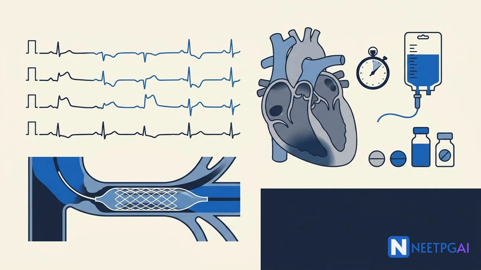
Clinical Case: 45-Year-Old Smoker with Crushing Chest Pain — Inferior STEMI Diagnosis, Reperfusion, and Complications for NEET PG
NEET PG inferior STEMI case: 45-yo male smoker, ECG ST elevation in II/III/aVF, V4R for RV involvement, primary PCI vs fibrinolysis, DAPT, Killip class, MCQ traps.
Version 1.0 — Published April 2026
Quick Answer
Acute STEMI is the highest-mortality cardiology diagnosis NEET PG tests, and inferior STEMI in a young Indian smoker is the most-tested vignette across recent papers. In a 45-year-old male smoker with crushing retrosternal chest pain radiating to the left arm, diaphoresis, and 2 hours of symptoms with ST elevation in II, III, and aVF, follow this 7-step workflow:
- Recognise the STEMI within 10 minutes — 12-lead ECG, ST elevation in two contiguous inferior leads
- Localize and assess RV involvement — ST elevation III > II suggests RCA; check V4R for RV infarct
- Initiate MONA-B carefully — aspirin 325 mg chewed, P2Y12 loading, anticoagulation, atorvastatin 80 mg; NO nitrates if RV involvement
- Reperfuse fast — primary PCI with door-to-balloon ≤90 min, or fibrinolysis with door-to-needle ≤30 min if PCI unavailable in 120 min
- Risk-stratify with Killip class — bedside grading drives ICU vs ward and IABP decisions
- Start guideline-directed medical therapy — DAPT, beta-blocker, ACEi/ARB, high-intensity statin, MRA if EF <40 with HF/DM
- Watch for complications — arrhythmias, mechanical (free-wall rupture, VSR, papillary muscle rupture), cardiogenic shock
Door-to-balloon time is the single most important variable: every 30-minute delay increases 1-year mortality by 7.5 percent.
The case
A 45-year-old marketing manager from Mumbai is brought to the emergency department by his wife at 7:45 AM with sudden onset crushing retrosternal chest pain that started about 2 hours ago while he was getting ready for work. He describes it as a heavy "elephant sitting on my chest," radiating to the left arm and jaw, associated with profuse sweating, nausea, and one episode of vomiting. He took a domperidone tablet thinking it was indigestion. The pain has not relented despite rest. He smokes 15 cigarettes per day for 20 years (15 pack-years), is a social drinker, and has a strong family history — his father died of a heart attack at age 52. He has hypertension diagnosed 3 years ago on telmisartan 40 mg, but is non-compliant. No diabetes, no prior cardiac event, no medication allergies.
On arrival, vitals are: pulse 56/min regular, BP 96/62 mmHg, respiratory rate 22/min, SpO2 95 percent on room air, temperature 36.6 C, capillary glucose 138 mg/dL. He is anxious, diaphoretic, in obvious distress, clutching his chest. Pain score 9/10. JVP appears slightly elevated. Carotid pulses normal in volume and contour. Apex beat is in the 5th intercostal space, mid-clavicular line, normal in character. On auscultation: heart sounds S1 and S2 normal, no S3, no S4, no murmurs, no pericardial rub. Lungs are clear bilaterally — no crackles, no wheeze. Abdomen soft, non-tender. No peripheral edema. Distal pulses 2+ bilaterally. Neurologically intact.
The triage nurse hands over the 12-lead ECG done within 7 minutes of arrival.
ECG findings: Sinus bradycardia at 56/min. ST elevation of 3 mm in lead III, 2 mm in lead II, and 2.5 mm in aVF with hyperacute T waves. Reciprocal ST depression of 2 mm in leads I and aVL. No ST elevation in V1-V6. No pathological Q waves yet. PR interval normal. QRS narrow.
The on-call cardiologist is paged immediately, the cath lab is activated, and the right-sided ECG is ordered.
ABCD assessment and initial investigations
Acute inferior STEMI in a 45-year-old smoker is a time-critical diagnosis where every minute of delayed reperfusion translates into infarcted myocardium. Resuscitation runs parallel to diagnostic workup — they are not sequential.
A — Airway: Patent. GCS 15. No airway compromise. Oxygen by nasal cannula at 2 L/min (SpO2 95 percent — give O2 only if SpO2 below 90; routine high-flow O2 in normoxic patients increases infarct size).
B — Breathing: RR 22, SpO2 95 on 2 L. CXR ordered to rule out aortic dissection (mediastinal widening) and pulmonary edema. Continuous SpO2 monitoring.
C — Circulation: BP 96/62 with bradycardia — concerning for inferior STEMI with right ventricular involvement and Bezold-Jarisch reflex (vagally-mediated bradycardia and hypotension common in RCA territory infarcts). Two large-bore peripheral IV lines (18G). Cautious IV fluid bolus of normal saline 250 mL with re-assessment — RV infarction patients are preload-dependent. Avoid nitrates until RV involvement is excluded. Telemetry continuous monitoring for arrhythmia. Pads on for defibrillation readiness.
D — Disability/Dextrose: GCS 15, glucose 138. Pain score 9/10 — IV morphine 2-4 mg titrated cautiously (aware that it slows P2Y12 absorption and may worsen hypotension).
Initial pharmacotherapy (within 10 minutes):
- Aspirin 325 mg chewed — irreversible platelet COX-1 inhibition; mortality benefit established (ISIS-2)
- Clopidogrel 600 mg loading OR ticagrelor 180 mg loading OR prasugrel 60 mg loading (P2Y12 receptor blockade; prasugrel only if PCI confirmed and no prior stroke/TIA, age <75, weight >60 kg)
- Atorvastatin 80 mg — mortality benefit independent of LDL reduction (PROVE-IT, ARMYDA)
- Unfractionated heparin 60 U/kg bolus (max 4000 U) followed by 12 U/kg/hr, OR enoxaparin 30 mg IV bolus + 1 mg/kg SC
- NO nitrates until RV involvement excluded (RV infarction is preload-dependent; nitrates can precipitate cardiac arrest)
- Beta-blocker — IV metoprolol 5 mg deferred in this patient given bradycardia and borderline BP; oral after stabilisation
Right-sided ECG (V4R): ST elevation of 1.5 mm in V4R confirms RV involvement. This changes management — fluid loading becomes the mainstay; nitrates and high-dose diuretics are contraindicated.
Initial labs (within 30 minutes):
- High-sensitivity troponin I: 145 ng/L on arrival (above 99th percentile URL of 26 ng/L) — rising trend at 3 hours to 1,820 ng/L confirms acute MI
- CBC: Hb 14.2 g/dL, WBC 11,200 (mild stress leukocytosis), platelets 256,000
- Renal: BUN 18, creatinine 1.0, eGFR 88 mL/min (important for contrast load planning)
- Electrolytes: Na 138, K 3.8, Mg 1.9 — replete K to >4 and Mg to >2 (anti-arrhythmic prophylaxis)
- Coagulation: PT/INR 1.0, aPTT 28 s (baseline before heparin)
- Lipid profile: LDL 148, HDL 32, TG 220 — atherogenic profile
- HbA1c: 5.6 percent (no diabetes)
- CXR: Normal cardiac silhouette, no pulmonary edema, no mediastinal widening
- Bedside echocardiogram (focused): Hypokinesia of inferior wall, RV free wall hypokinesia, LVEF estimated 50 percent, no pericardial effusion, no obvious mechanical complication
The diagnostic algorithm — confirming STEMI
NEET PG tests the 4th Universal Definition of MI explicitly. STEMI diagnosis requires:
STEMI ECG criteria
| Lead group | ST elevation threshold |
|---|---|
| V2-V3 (men ≥40 years) | ≥2 mm at the J-point |
| V2-V3 (men <40 years) | ≥2.5 mm at the J-point |
| V2-V3 (women, all ages) | ≥1.5 mm at the J-point |
| All other leads (men and women) | ≥1 mm in two contiguous leads |
| Posterior leads V7-V9 | ≥0.5 mm |
| Right-sided V3R-V4R | ≥0.5 mm (≥1 mm in men <30) |
Plus ischemic symptoms or new LBBB equivalent (Sgarbossa criteria). Troponin is not required for STEMI diagnosis at presentation — reperfusion should not wait for biomarker confirmation.
Localizing the culprit artery
| Leads with ST elevation | Likely culprit |
|---|---|
| II, III, aVF | RCA (80%) or LCx (20%) |
| V1-V4 | Proximal LAD |
| V5-V6, I, aVL | LCx or distal LAD/diagonal |
| V1-V6 + I + aVL | Proximal LAD ("widow-maker") |
| ST depression V1-V3 with tall R waves | Posterior MI (LCx or RCA) — confirm with V7-V9 |
| ST elevation aVR > ST elevation V1, with diffuse ST depression | Left main or 3-vessel disease |
For our patient: III > II elevation, reciprocal depression in I/aVL, RV involvement on V4R — RCA proximal occlusion with right ventricular branch involvement.
Troponin kinetics
| Marker | Rise | Peak | Return to baseline |
|---|---|---|---|
| High-sensitivity troponin I/T | 1-3 hr | 12-24 hr | 7-14 days |
| CK-MB | 3-6 hr | 12-24 hr | 48-72 hr |
| Myoglobin (rarely used now) | 1-2 hr | 6-12 hr | 24 hr |
| LDH (historical) | 24-48 hr | 3-6 days | 8-14 days |
CK-MB is the only marker useful for diagnosing re-infarction within the first 14 days because troponin remains elevated. Modern protocols use 0/1-hour or 0/3-hour high-sensitivity troponin algorithms for non-STEMI rule-out.
Diagnosis
Acute inferior wall ST-elevation myocardial infarction (STEMI) with right ventricular involvement, presumed RCA proximal occlusion, Killip Class I, presenting 2 hours from symptom onset in a 45-year-old male smoker with hypertension, dyslipidemia, and strong family history — Class I indication for emergent primary percutaneous coronary intervention.
Differential diagnosis of acute chest pain
Running the differential matters because misdiagnosed STEMI mimics carry catastrophic management implications.
Cannot-miss diagnoses
- STEMI — ST elevation pattern, ischemic risk factors, troponin rise
- NSTEMI / unstable angina — ischemic pain without ST elevation, troponin may rise (NSTEMI) or not (UA)
- Aortic dissection — tearing chest/back pain, BP differential between arms >20 mmHg, mediastinal widening on CXR, false lumen on CT angiogram. CRITICAL: thrombolysis or DAPT in undiagnosed dissection is fatal
- Pulmonary embolism — pleuritic pain, hypoxia, tachycardia, S1Q3T3 pattern, raised D-dimer, RV strain on echo
- Tension pneumothorax — sudden dyspnea, tracheal deviation, absent breath sounds, hyperresonance
- Cardiac tamponade — Beck triad (hypotension, raised JVP, muffled heart sounds), pulsus paradoxus, electrical alternans
- Esophageal rupture (Boerhaave) — vomiting then severe chest pain, mediastinal air on CXR
- Acute pericarditis — pleuritic positional pain, friction rub, diffuse ST elevation with PR depression
Why our patient's pain is STEMI
Five features lock it in: (1) classic ischemic character (crushing, retrosternal, radiating to left arm and jaw, with diaphoresis and nausea), (2) persistent for over 20 minutes (rules out stable angina), (3) diagnostic ECG with ST elevation in contiguous inferior leads with reciprocal changes, (4) risk profile (smoker, hypertensive, dyslipidemic, strong family history), and (5) rising troponin trajectory. The reciprocal ST depression in I and aVL is particularly helpful — it reduces the chance of pericarditis (which has diffuse ST elevation without reciprocal changes) and early repolarization (which has no reciprocal changes).
Management — door-to-balloon ≤90 minutes is the goal
Reperfusion strategy decision tree
Primary PCI (Class I, Level of Evidence A) is preferred when:
- First-medical-contact-to-balloon time ≤90 minutes at a PCI-capable hospital
- Transfer-to-balloon time ≤120 minutes from a non-PCI hospital
- Cardiogenic shock or severe HF (Killip III-IV) regardless of time
- Contraindications to fibrinolysis
- Diagnostic uncertainty (helpful angiography)
- Late presentation >3 hours from symptom onset
Fibrinolysis is acceptable when:
- PCI cannot be delivered within 120 minutes
- Symptom onset within 3 hours
- No contraindications
- Door-to-needle target ≤30 minutes
- All fibrinolytic patients must transfer to PCI-capable centre for rescue PCI if reperfusion fails (persistent ST elevation, ongoing pain at 60-90 min) or routine PCI within 24 hours if successful (PRAGUE-2, CARESS-in-AMI, TRANSFER-AMI)
Fibrinolytic agents
| Agent | Class | Dose | Notes |
|---|---|---|---|
| Tenecteplase (TNK-tPA) | Fibrin-specific | Single weight-adjusted IV bolus (30-50 mg) | Easiest to administer; preferred in India |
| Alteplase (rt-PA) | Fibrin-specific | 15 mg bolus, then 0.75 mg/kg over 30 min, then 0.5 mg/kg over 60 min (max 100 mg) | More complex regimen |
| Reteplase | Fibrin-specific | Two 10 U bolus 30 min apart | Less common in India |
| Streptokinase | Non-fibrin-specific | 1.5 million U over 30-60 min | Cheap; antigenic — cannot repeat within 6 months; widely available in India |
Absolute contraindications to fibrinolysis (NEET PG favourite)
- Any prior intracranial hemorrhage
- Known structural cerebrovascular lesion (AVM)
- Known malignant intracranial neoplasm
- Ischemic stroke within 3 months (except acute ischemic stroke within 4.5 hr — that gets thrombolysis itself)
- Suspected aortic dissection
- Active bleeding or bleeding diathesis (excluding menses)
- Significant closed-head or facial trauma within 3 months
- Intracranial or spinal surgery within 2 months
- Severe uncontrolled hypertension >180/110 unresponsive to therapy
- For streptokinase: prior streptokinase use within 6 months
Relative contraindications: active peptic ulcer, recent prolonged CPR >10 min, current anticoagulation (INR >1.7), pregnancy, recent major surgery within 3 weeks, chronic poorly-controlled severe hypertension, dementia or known intracranial pathology.
Our patient's pathway
Mumbai with a PCI-capable centre 25 minutes away. The decision is straightforward: transfer to cath lab for primary PCI. While transferring:
- Aspirin 325 mg chewed (already given)
- Ticagrelor 180 mg PO (preferred over clopidogrel in PCI; avoid prasugrel in stroke history or age ≥75)
- UFH 60 U/kg bolus, infusion 12 U/kg/hr
- Cautious 250 mL NS bolus for RV preload (BP improved to 110/70)
- NO nitrates (RV involvement)
- Atorvastatin 80 mg
- Pain control with low-dose IV morphine
- Continuous telemetry; defibrillator pads on
Cath findings: Total occlusion of proximal RCA before the RV branch; mid-LAD 40 percent non-obstructive lesion; LCx unobstructed. DES (drug-eluting stent) deployed in RCA with TIMI 3 flow restored. Door-to-balloon time: 78 minutes. Post-procedure echo: LVEF 48 percent, residual inferior wall hypokinesia, mild TR.
Post-revascularization care — the GDMT bundle
Dual antiplatelet therapy (DAPT) for 12 months:
- Aspirin 75-100 mg daily lifelong
- Ticagrelor 90 mg BID for 12 months (or clopidogrel 75 mg daily if ticagrelor unavailable / poorly tolerated)
Beta-blocker within 24 hours if no contraindications (HF, hypotension, bradycardia, AV block):
- Metoprolol succinate 25-50 mg daily, uptitrated
- Carvedilol 6.25 mg BID, uptitrated
- Mortality benefit (CAPRICORN, ISIS-1)
ACE inhibitor / ARB within 24 hours, especially if anterior MI, EF <40, HF, DM:
- Ramipril 2.5 mg BID uptitrated to 5 mg BID
- Telmisartan 40 mg daily if ACE-I cough
High-intensity statin (atorvastatin 80 mg or rosuvastatin 20-40 mg) — not LDL-driven; mortality benefit independent of lipid lowering
Mineralocorticoid receptor antagonist (eplerenone 25-50 mg or spironolactone 25 mg) if EF ≤40 with HF or DM (EPHESUS)
Cardiac rehabilitation, smoking cessation (varenicline + counseling), Mediterranean diet, weight loss, BP control to <130/80, LDL target <55 mg/dL post-MI (2019 ESC, 2022 AHA)
Complications — what kills these patients
Early complications (first 24-48 hours)
Arrhythmias (commonest, contribute most to early mortality)
- Ventricular fibrillation / pulseless VT — primary VF (within 48 hr) does not affect long-term prognosis if defibrillated; secondary VF (after 48 hr in HF context) signals worse prognosis
- Sustained monomorphic VT — from scar reentry; requires synchronised cardioversion
- Accelerated idioventricular rhythm — common reperfusion arrhythmia, benign, no treatment needed
- AV blocks in inferior MI — common (RCA supplies AV node in 90 percent); usually transient; atropine, temporary pacing if symptomatic; permanent pacemaker rarely needed
- AV blocks in anterior MI — rare but ominous (extensive infarct involving conduction system); often permanent; PPM commonly required
- Sinus bradycardia / Bezold-Jarisch reflex — vagal reflex from inferior MI; atropine, fluids; usually resolves
Pump failure
- Cardiogenic shock (Killip IV) — mortality 60-80 percent; treat with revascularization, IABP, inotropes (norepinephrine, dobutamine), VA-ECMO or Impella in selected centres
- Acute pulmonary edema (Killip III) — diuretics, IV nitrates (avoid in RV infarct), non-invasive ventilation, urgent revascularization
Mechanical complications (3-7 days, but can occur earlier)
| Complication | Timing | Murmur / sign | Management |
|---|---|---|---|
| Free-wall rupture | Day 2-7 | Sudden tamponade, PEA arrest | Emergent surgical repair; almost always fatal |
| Ventricular septal rupture | Day 3-7 | New harsh holosystolic murmur at LSB, thrill | Diuretics, IABP, urgent surgical or percutaneous closure |
| Papillary muscle rupture | Day 2-7 | Acute MR — soft systolic murmur, flash pulmonary edema (often no audible murmur from rapid LA pressure equilibration) | IABP, afterload reduction, urgent MV repair/replacement |
| LV pseudoaneurysm | Days to weeks | Persistent ST elevation, contained rupture | Surgical repair |
| True LV aneurysm | Weeks to months | Persistent ST elevation, double impulse, S3, mural thrombus | ACE-I, anticoagulation if thrombus, rarely surgical |
| Dressler syndrome | 2-10 weeks | Pleuropericarditis, fever, pleural effusion (immune-mediated) | NSAIDs, colchicine; avoid corticosteroids early |
Other complications
- LV thrombus — anterior MI; warfarin or DOAC for 3-6 months
- Atrial fibrillation — ~10 percent post-MI; rate/rhythm control + anticoagulation per CHA2DS2-VASc
- Right ventricular infarction — preload-dependent; fluid loading; avoid nitrates and high-dose diuretics
- Recurrent ischemia / re-infarction — angiography, additional revascularization; CK-MB more useful than troponin for rediagnosis
How NEET PG tests acute MI
NEET PG tests this topic through seven recurring patterns. Recognise the pattern and the question collapses to a 30-second answer.
Pattern 1 — The ECG-localization question: Vignette gives ST elevation in II, III, aVF with ST depression in I and aVL. Culprit artery? RCA in 80 percent (LCx in 20 percent if II > III with ST depression in V1-V3). Trap: answers offering "LAD" — that produces precordial ST elevation, not inferior.
Pattern 2 — The RV-involvement question: Inferior STEMI with hypotension, clear lungs, and raised JVP. Next investigation? Right-sided ECG (V4R). Best initial management of confirmed RV infarction? IV fluid loading; AVOID nitrates and morphine. Trap: answers offering "IV nitroglycerin for chest pain" — can precipitate cardiac arrest in preload-dependent RV infarct.
Pattern 3 — The reperfusion-strategy question: STEMI patient at non-PCI hospital, transfer-to-balloon estimated 90 minutes. Best management? Transfer for primary PCI. Same scenario but transfer-to-balloon 150 minutes? Fibrinolysis on-site, then transfer for routine PCI within 24 hours. Trap: answers offering "wait for cardiologist consultation" — delay kills myocardium.
Pattern 4 — The contraindication question: STEMI presenting 2 hours after onset, recent ischemic stroke 6 weeks ago. Reperfusion? Primary PCI (fibrinolysis contraindicated within 3 months of ischemic stroke). Trap: answers offering "tenecteplase" — absolute contraindication.
Pattern 5 — The mechanical-complication question: Day-5 post-MI patient with new harsh holosystolic murmur at left sternal border and acute pulmonary edema. Diagnosis? Ventricular septal rupture. Acute MR from papillary muscle rupture often has NO audible murmur because of rapid LA pressure equilibration — beware this trap.
Pattern 6 — The Killip-class question: Post-MI patient with bibasal crackles up to mid-zones and S3. Killip class? III (pulmonary edema). Mortality? ~38 percent. Class IV is cardiogenic shock with mortality 60-80 percent.
Pattern 7 — The medication-after-MI question: Best antiplatelet duration after PCI for STEMI? DAPT for 12 months, then aspirin lifelong. Statin choice? High-intensity (atorvastatin 80 mg or rosuvastatin 20-40 mg) regardless of baseline LDL. Beta-blocker in HFrEF post-MI? Carvedilol, metoprolol succinate, or bisoprolol — mortality benefit.
High-yield one-liners:
- STEMI = ST elevation ≥1 mm in 2 contiguous leads (≥2 mm V2-V3 men ≥40)
- Door-to-balloon ≤90 min, door-to-needle ≤30 min
- Inferior STEMI: III > II = RCA; II > III with I/aVL ST depression = LCx
- RV infarction: V4R ST elevation, preload-dependent, avoid nitrates and morphine
- DAPT 12 months post-PCI; aspirin lifelong; high-intensity statin always
- Free-wall rupture day 2-7, fatal; VSR new murmur; papillary muscle rupture acute MR often without murmur
- Killip I = no HF (6%); II = crackles/S3 (17%); III = pulmonary edema (38%); IV = shock (60-80%)
- CK-MB best for re-infarction within 14 days (troponin still elevated)
- Dressler syndrome 2-10 weeks post-MI; immune-mediated pleuropericarditis
- Streptokinase: cannot repeat within 6 months (antigenic)
Frequently Asked Questions
How is the universal definition of myocardial infarction applied at the bedside?
The 4th Universal Definition of MI requires a rise and/or fall of cardiac troponin with at least one value above the 99th percentile upper reference limit, plus at least one of: ischemic symptoms, new ischemic ECG changes (ST elevation, ST depression, T inversion, new LBBB), pathological Q waves, imaging evidence of new loss of viable myocardium or regional wall-motion abnormality, or identification of an intracoronary thrombus on angiography or autopsy. STEMI is defined separately by ischemic symptoms plus persistent ST elevation greater than 1 mm in two contiguous leads (greater than 2 mm in V2-V3 in men over 40, greater than 1.5 mm in women) or new LBBB — and triggers immediate reperfusion without waiting for troponin.
What ECG features localize an inferior STEMI and identify right ventricular involvement?
Inferior STEMI shows ST elevation in leads II, III, and aVF, often with reciprocal ST depression in I and aVL. ST elevation greater in lead III than lead II suggests an RCA culprit (the more common scenario, ~80 percent), while ST elevation greater in II than III with ST depression in I and aVL suggests left circumflex involvement. Right ventricular involvement is suggested by ST elevation in V1 and confirmed by ST elevation greater than 1 mm in V4R on a right-sided ECG. RV infarction occurs in 30-50 percent of inferior STEMIs and changes management — patients become preload-dependent, so nitrates are contraindicated and IV fluid loading is the cornerstone.
When is primary PCI preferred over fibrinolysis in STEMI?
Primary PCI is preferred whenever it can be delivered with a first-medical-contact-to-balloon time within 90 minutes (or 120 minutes if transferring from a non-PCI hospital). PCI is also preferred regardless of time in cardiogenic shock, severe heart failure (Killip III-IV), contraindications to fibrinolysis, late presentation (over 3 hours from symptom onset), and diagnostic uncertainty. Fibrinolysis is acceptable when PCI is unavailable within 120 minutes, presentation is within 3 hours of symptom onset, and there are no contraindications. Door-to-needle target is 30 minutes; door-to-balloon target is 90 minutes.
What are the absolute contraindications to fibrinolytic therapy in STEMI?
Absolute contraindications: any prior intracranial hemorrhage; known structural cerebrovascular lesion (AVM); known malignant intracranial neoplasm; ischemic stroke within 3 months (except acute ischemic stroke within 4.5 hours); suspected aortic dissection; active bleeding or bleeding diathesis (excluding menses); significant closed-head or facial trauma within 3 months; intracranial or intraspinal surgery within 2 months; severe uncontrolled hypertension over 180/110 unresponsive to therapy; for streptokinase, prior streptokinase use within 6 months. Relative contraindications include active peptic ulcer, recent prolonged CPR, current anticoagulation, pregnancy, recent major surgery within 3 weeks, and chronic severe poorly-controlled hypertension.
What is the Killip classification and how does it predict mortality in STEMI?
The Killip classification grades acute heart failure in STEMI based on bedside examination: Class I — no clinical signs of heart failure (in-hospital mortality ~6 percent); Class II — basal lung crackles, S3 gallop, or elevated JVP indicating mild-to-moderate failure (mortality ~17 percent); Class III — frank pulmonary edema with crackles in over half the lung fields (mortality ~38 percent); Class IV — cardiogenic shock with hypotension under 90 mmHg systolic and peripheral hypoperfusion (mortality 60-80 percent without revascularization). Killip class is a Class I indication for risk stratification and a major driver of decisions about IABP, mechanical support, and ICU admission.
This content is for educational purposes for NEET PG exam preparation. It is not a substitute for professional medical advice, diagnosis, or treatment. Clinical information has been reviewed by qualified medical professionals.
Written by: NEETPGAI Editorial Team Reviewed by: Pending SME Review Last reviewed: April 2026
This content is for educational purposes for NEET PG exam preparation. It is not a substitute for professional medical advice, diagnosis, or treatment. Clinical information has been reviewed by qualified medical professionals.
Ready to put this into practice?
Start practicing NEET PG MCQs with AI-powered explanations.
Start Free PracticeYour Next Step
Related Study Guides
Wilms Tumor and Neuroblastoma Pediatric Oncology for NEET PG 2026
Master Wilms tumour vs neuroblastoma, WAGR/Denys-Drash/Beckwith-Wiedemann, NMYC amplification, NWTS/INRG staging and treatment for NEET PG 2026.
Otitis Media and Sinusitis ENT Guide for NEET PG 2026
Master AOM, OME, CSOM tubotympanic vs atticoantral, cholesteatoma, FESS indications and post-COVID mucormycosis sinusitis for NEET PG 2026.
Image MCQ: Pediatric Imaging Findings for NEET PG (Intussusception, Pyloric Stenosis, Hirschsprung, VUR, DDH)
5 pediatric imaging MCQs for NEET PG: intussusception target sign, hypertrophic pyloric stenosis string sign, Hirschsprung contrast enema, VUR grading on MCUG, DDH Graf alpha angle.
Join our NEET PG community
Daily MCQs, study tips, and topper strategies on Telegram.
Join on Telegram →